수근관절 불안정성(Carpal instability) : 손목에 외상을 입은 뒤 손목이 불안정한 느낌이 있어요

수근관절 불안정성의 정의
손목의 외재 인대(extrinsic ligament : radiocarpal)와 내재 인대(intrinsic ligaments : intercarpal)는 손목 관절의 안정성을 유지시켜 주는데 손목의 불안정성은 이 인대들의 손상으로 인해 발생한 불안정성을 말합니다.
가장 흔한 원인은 주상골의 불안정 골절, 주상골-월상골간 해리(scapholunate dissociation), 월상골-삼각골간 해리(lunotriquetral dissociation)입니다.
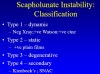
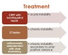
수근관절 불안정성의 분류
- CID(Carpal instability dissociative) : 같은 열(same carpal row)에 위치한 수근골들 또는 수근골들 사이의 이상입니다.
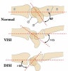
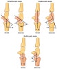
1) Scapholunate dissociation(=DISI : Dorsal intercalated segmental instability)
Scapholunate interosseous ligament의 손상이 주 원인으로 월상골이 배측으로 향해 신전하고, 주상골은 수장쪽으로 굴곡합니다.
병력은 acute FOOSH injury가 있거나 degenerative rupture입니다.
손상정도와 손상의 인대의 위치에 따라 predynamic -> dynamic -> static instability -> SLAC으로 진행합니다.
임상증상
요골 배측의 손목 통증이 있고 쥐는 힘의 감소를 호소합니다.
Wrist hyperextension, radial deviation시 통증이 가중됩니다.
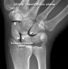
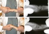
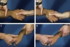
Scaphoid Shift test(=Watson test)
진단
X-ray를 촬영합니다(PA view, lateral view, ulnar deviation and radial deviation view, flexion and extension view).
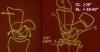
Scapholunate angle>60º, Radiolunate angle>15º를 보입니다.
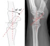
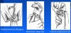
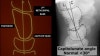
* Scapholunate angle
Normal : 30 - 60°
Questionably abnormal : 60 - 80°
Abnormal : > 80° indicates instability of the wrist.
* Scaphoid axis
The line through the midpoints of its proximal and distal poles.
Since the midpoint of the proximal pole is often difficult to appreciate, an almost parallel line can be used that is traced along the most ventral points of the proximal and distal poles of the bone.
* Lunate axis
The axis of the lunate runs through the midpoints of the convex proximal and concave distal joint surfaces and can best be drawn by finding the perpendicular to a line joining the distal palmar and dorsal borders of the bone as demonstrated on the left.
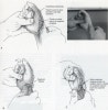
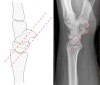
* Normal scapholunate space : The normal scapholunate space(arrowheads) is similar in width to other normal intercarpal joints(arrows).
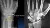
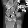
* Scapholunate widening
Widening (arrowheads) of the scapholunate distance > 2 mm
The space is obviously wider than the other intercarpal spaces (arrows).
This results in the 'Terry Thomas sign'.
Widening if the scapholunate space indicates a tear injury of the scapholunate ligament.
- Lunotriquetral dissocation(=VISI : Volar intercalated segmental instability)
두번째로 흔하기는 하나 그 빈도가 높지는 않습니다.
Lunotriquetral ligament의 점진적인 손상과 관련된 형태로 lunotriquetral ligament의 파열, 주상골와 함께 월상골도 수장쪽으로 굴곡합니다.
Lunotriquetral ligament의 단독손상이 대부분이고, 주변의 외인성인대와 함께 손상되어야 비로소 Lunotriquetral instability가 발생하게 되며, TFCC 병변이 동반되는 경우도 빈번합니다.

임상증상
척골 측의 손목 통증이 있습니다.
Ulnar deviation시 통증이 가중됩니다.
진단
X-ray를 촬영합니다(PA view, lateral view, ulnar deviation and radial deviation view, flexion and extension view).
Scapholunate angle<30º, Radiolunate angle<15º를 보입니다.
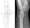
Cf) Capitolunate instability
* Capitolunate angle
The capitate axis joins the midportion of the proximal convexity of the third metacarpal and that of the proximal surface of the capitate.
Capitolunate angle > 30º indicates instability of the wrist.
- CIND(Carpal instability non-dissociative) : 열과 열 사이, 즉 요골과 근위열 수근골 사이, 수근골의 근위열과 원위열 사이 혹은 두 군데 모두에 이상이 있어 기능이상(dysfunction)이 온 경우입니다.
1) Radiocarpal CIND : High energy trauma, 관절질환, developmental anomaly(Madelung’s deformity), 원위부 요골절제 같은 수술적 치료후에 발생합니다.
요골과 척골에 대한 수근골들의 상대적인 이동에 따라 ulnar, dorsal, volar, combined translocation으로 구분합니다.

2) Midcarpal CIND : Triquetro-hamate-capitate ligament와 scaphocapitate, scaphotrapezio-trapezoid ligament의 손상에 의합니다(외상의 병력은 없습니다).
Palmar midcarpal instability, Dorsal midcarpal instability, Dorsal and palmar instability, Extrinsic midcarpal instability으로 분류할 수 있습니다.
임상증상
아탈구가 있으며 통증은 있기도 없기도 합니다.
손목에 힘 빠짐(giving way)을 호소합니다.
Clunking sign를 호소합니다("clunk" when wrist is moved ulnarly from flexion to extension with an axial load).
이학적 검사
Generalized ligamentous laxity를 보입니다.
영상 검사
X-ray를 촬영합니다(PA view, lateral view, ulnar deviation and radial deviation view, flexion and extension view).
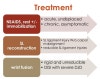
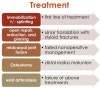
치료
1차적으로 splint를 적용하여 움직임을 제한합니다.
보존적 치료로 호전되지 않으면 여러가지 수술방법을 고려합니다.
치유과정에서 진통, 인대 유합의 촉진을 위해 침 치료를 적용합니다(근위축 예방과 근육 운동시의 동통완화 효과를 목적으로 합니다).
-
CIC(Carpal instability complex) : Perilunate dislocation
-
CIA(Carpal instability adaptive) : 원위 요골 골절에서 관절면이 정상과 반대로 후방 경사로 된 경우가 대부분으로 월상골은 신전되는 DISI 양상을 보입니다.
참고문헌
- 정형외과 진료편람. 서울대학교 의과대학 정형외과학 교실. 2013
2. 쉽게 배우는 정형외과. Okada kyoji. 2014
-
일차진료의를 위한 정형외과 진단과 치료. 김지형. 2011
-
AAOS 핵심 정형외과학 4판. John F. Sarwark. 2013
-
임상의를 위한 통증의 영상진단과 치료. Steven D. Waldman. 2012
-
응급영상진단의 ABC. Otto Chan. 2010
-
스포츠 침구임상 매뉴얼. Matsumoto tadasu. 2007