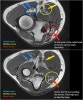
팔꿈치 관절의 Basic MRI
1. Ulnar Collateral Ligament
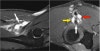
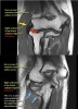
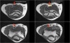
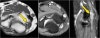
연속적인 관상면 영상에서 UCL을 볼 수 있습니다.
정상적인 UCL이 sublime tubercle에 확실하게 붙어 있는 것을 볼 수 있습니다.
Proximal part에서 약간의 high signal이 보이는 것은 정상입니다(arrow).
2. Lateral Collateral Ligament
3. Common Extensor Tendon
The common extensor tendon originates at the lateral epicondyle. On a T1W-images the tendon should have a low signal intensity (yellow arrow).
4. Common Flexor Tendon
The common flexor tendon originates at the medial epicondyle.
On a T1W-images the tendon should have a low signal intensity (red arrow).
5. Brachialis tendon
On a sagittal view, when you compare the brachialis tendon (yellow arrows) with the biceps tendon (red arrows), notice that the brachialis is almost all muscle. It only has a very short tendon distally.
6. Biceps tendon
7. Nerves
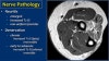
1) Pathology
This is a nice example of subacute denervation.
Notice on the T1W-image that there is no atrophy. Only edema on the T2W-image.
This was due to proximal radial neuropathy.
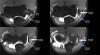
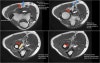
- Ulnar nerve
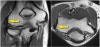
Here we see the ulnar nerve within the cubital tunnel.
The posterior band of the ulnar collateral band forms the floor of the tunnel, while the retinaculum forms the roof.
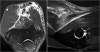
* Ulnar neuritis
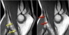
There is enlargement of the nerve. On the T2W-image there is high signal.
Another sign is non-uniform enlargement of the fascicles, which is seen on the sagittal image (arrow).
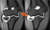
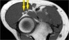
- Radial nerve
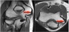
The radial nerve can be best identified at the level of the radial head, where you can see superficial and deep branches in the radial tunnel (arrows). This is a very consistent place to find the radial nerve.
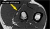
* Arcade of Frohse
The deep radial branches form the posterior interosseus nerve which penetrates the supinator muscle at the arcade of Frohse (arrow).
4) Median nerve
The median nerve goes down behind the Lacertus fibrosis, which is the aponeurosis of the biceps and penetrates the pronator muscle.
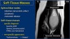
8. Soft Tissue Masses
On MR a mass was seen just above the medial epicondyle, The mass is very heterogeneous as is the enhancement.
Based on the MR-findings you still have to call this mass indeterminate.
The final diagnosis was cat scratch disease based on high Bartonella henselae titers.
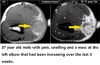
It looks homogeneous and cystic. Continue with the post-Gd image. The final diagnosis at biopsy was Lymphoma.
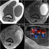
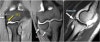
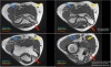
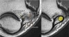
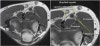
* Radiobicipital bursitis
Bursa(white arrow in left)
Notice that the biceps is intact. Next to the radiobicipital bursa (yellow arrow), also an interosseous bursa (red arrow)
Sometimes these masses mimic a tumor or they can cause impingement on the radial nerve when they become very large.