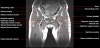
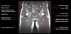
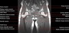
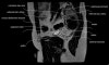
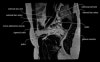
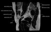
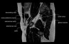
엉덩이 관절(고관절)의 MRI 단면 영상
- Checklist
1) Femur : Osteonecrosis, Fracture or Edema
2) Cartilage surface : Fissure, Fraying, Thinning or Defect
3) Joint recess : Chondral debris or Corpora aliena
4) Labrum : Tear, Detachment, Fraying or Degeneration
5) Acetabulum : Shallow contour?
6) Muscle and tendon : Tear or Strain
7) Trochanteric or Iliopsoas bursitis?
8) Check the symphysis pubis, superior/inferior pubic rami, ilium, sacroiliac joint and sacrum on large FOV coronal images
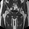
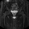
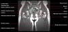
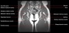
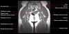
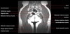
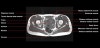
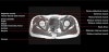
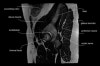
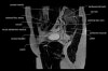
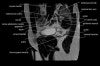
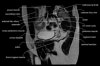
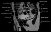
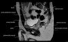
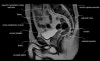
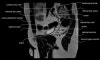
- Coronal section
T1 Allows for evaluation of both hips simultaneously even though the patient may be symptomatic in only one hip.
T2 allows for detection of abnormal fluid in both hips which may be seen in avascular necrosis, stress fractures, muscle tears or perilabral cysts.
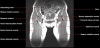
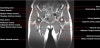
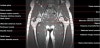
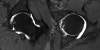
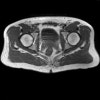
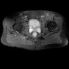
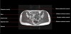
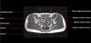
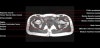
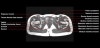
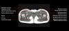
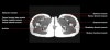
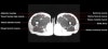
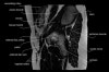
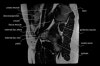
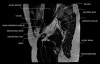
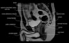
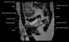
- Axial section
Axial T1 large field of view allows for evaluation of both hips simultaneously, particularly the acetabulae.
Axial PD fat suppressed is sensitive to fluid that may be present with avascular necrosis or stress fractures, while maintaining high a SNR.
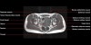
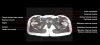
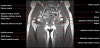
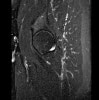
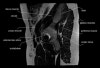
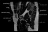
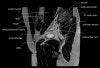
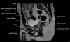
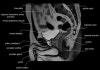
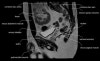
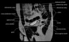
- Sagittal section
Sagittal PD fat suppression is sensitive to fluid that may be present with AVN or stress fractures.

5. Hip MR Arthrogram