안면 골절(Facial bone fractures)
1. Most common fractures
-
Isolated zygomatic arch fractures
-
‘Tripod' fractures
-
Orbital 'blowout' fractures
- Classification
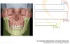
-
Upper 1/3 : Above the eyebrows -> Involves frontal sinuses & supraorbital ridges
-
Middle 1/3 : Above the mouth -> Central, Lateral
-
Lower 1/3 : Mandible
3. Mandible Fracture
- 개요
(1) 얼굴을 아래쪽으로 중간 정도 이상의 에너지가 전달될 때 발생한다.
(2) 외력에 취약한 지점, 또는 질병이 있는 부위에 골절이 잘 발생한다.
(3) 골반과 유사한 원형의 구조물로 만약 하나의 골절을 발견했다면, 다른 골절이 있는지 주의 깊게 살펴봐야 한다.
(4) Condylar fx와 TMJ injury는 구분이 어려울 수 있으므로 다른 영상이 필수적이다.
- Symptom
(1) 교합의 주관적인 변화, 손상의 개구장애
(2) 통증, 부종, 이상감각 또는 alveolar nerve의 무감각
- Sign
(1) 이상교합, 뼈의 통증, 이상감각, 마찰음
(2) 귀의 출혈, 혀밑의 출혈
- Radiology
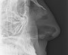
(1) This image shows an irregular fracture line passing across the mandible on the left. Careful inspection of the mandibular outline shows a second fracture at the mental symphysis.
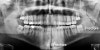
(2) Both fractures are again seen on the mandibular view.
4. Middle 1/3 facial fracture
| • Central - Nasal bone - Nasoethmoid - Maxilla : Lefort Ⅰ, Ⅱ, Ⅲ • Lateral : Zygomatic bone |
- Nasal fracture
(1) Most common site of facial fx : 다수가 복합골절의 양상, 응급의학 측면에서 매우 중요한 영역은 아니다.
(2) Frontal blow, Lateral blow, Blow from below
(3) 증상
① 코피, 복시, 부종
② Infraorbital nerve의 마비 및 감각 이상, 결막하 출혈
③ 부정교합, 안면의 비대칭, 치아의 손상
(4) 임상진단
① X-ray missed up to half
② When isolated, XR may be adequate
③ X-ray views : Laterals and Water
④ CT when concern more than mere nasal fx
2) Naso-Orbital-Ethmoidal(NOE) fracture
| (1) Fracture disrupting : Medial orbit + Nose + Ethmoid sinus (2) Medial canthal tendon slings globe to medial orbital wall (3) In NOE fracture, the tendon pulls fragment laterally causing telecanthus |
3) Le Fort fracture
| • Among the most severe facial fx • Progressively severe category from Ⅰ->Ⅲ • Separation(partial or complete) of maxilla from remainder face • All extend through posterior face transecting pterygoid plates • Ⅰ, Ⅱ, Ⅲ and combined |
(1) Le Fort Ⅰ fracture
| • Alveolar process 상부의 골절이 maxilla를 가로질러 분리된 상태 • 임상적으로 코뼈가 상부치아로부터 분리된다. |
(2) Le Fort Ⅱ fracture
| • 골절선이 코를 넘어가며 위쪽 치아와 코뼈가 하나의 덩어리로 분리된다. |
(3) Le Fort Ⅲ fracture
| • 안면부가 머리로 부터 완전 분리된다 (craniofacial dissociation). |
| * Pteryogoid plate fracture | |
| • 90-100% Le Foft fx • Isolated pterygoid plate fx very rare • Absence of pterygoid plate fx rules out Le Fort |
4) Isolated zygomatic arch fracture
| • Disruption of the middle McGrigor-Campbell line is due to a comminuted fracture of the right zygomatic arch • Following the upper and lower lines shows no fracture | |
| • Look for the 'elephant's trunk' appearance of the zygomatic arch. • Comparing the symptomatic side with the asymptomatic side can help reveal an abnormal contour of the zygomatic arch. | |
| • The zygomatic arch fracture is more easily seen on the OM30 (Occipito-Mental 30°) image. • On the left (the non-injured side) overlying structures give the impression of a fracture. However, careful scrutiny shows the cortex is intact. |
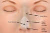
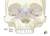
5) Tripod fracture
| • 삼각골절이란 이름은 광대뼈의 이마돌기(frontal process), 광대활, 위턱돌기(maxillary process)를 침범하기 때문에 붙여진 것이다. • 안면부 골절에서 가장 흔하다. • 뺨의 충격이나 광대뼈의 직접 손상에 의해 발생한다. • 진단은 Occipitomental 또는 Occipitomental 30º 영상에서 쉽게 진단되다. • 연부조직의 부종, 공기액체층, 일부의 회전(rotation)이 관찰된다(CT에 가장 잘 관찰). |
| • 1 : The zygoma (asterisk) is separated from the frontal bone at the zygomatico-frontal suture • 2 : Comminuted fracture of the zygomatic arch • 3 : Orbital floor fracture • 4 : Breach of the lateral wall of the maxillary antrum | |
| • The zygomatico-frontal suture (asterisk) appears relatively normal on this image and looks similar to the contralateral side. • Note : A slightly wide zygomatico-frontal suture should not be taken as significant unless accompanied by other evidence of injury on the same side. | |
| • A 'tripod' fracture has 4 visible components - not always all visible. • 1 : Orbital floor fracture • 2 : Fracture of the lateral wall of the maxillary antrum • 3 : Zygomatic arch fracture of the maxillary antrum is due to it filling with blood. • 4 : Widening of the zygomatico-frontal suture Increased density of the maxillary antrum is due to it filling with blood. | |
| • A fluid level of blood seen in the maxillary antrum may be the only obvious sign of fracture • A : Widened zygomatico-frontal suture • B : Zygomatic arch fracture • C : Orbital floor fracture • D : Lateral maxillary antrum wall fracture | |
| • The eye is drawn to the dark irregular line passing across the orbit which is the normal coronal suture • A systematic approach reveals a tripod injury with a large fracture of the orbital floor |
6) Orbital injury
(1) 주 증상과 징후는 얼룩출혈, 결막하출혈, 복시, 눈알함몰, Infraorbital 부위의 무감각 또는 감각이상 등
(2) 분류 : Orbital rim, Orbital floor, Medial wall, Lateral wall
(3) Orbital 'blowout' fracture
| • 안구의 직접적인 힘에 의해 발생하며, 이 힘에 의해 눈확의 바닥이나 안쪽 면이 골절되는 것을 말한다. • 즉각적인 수술의뢰가 필요 • On the left a 'teardrop' of soft tissue has herniated from the orbit into the maxillary antrum | |
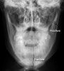
(4) Orbital emphysema
Occasionally a 'tripod' or 'blowout' fracture will cause a leak of air from the maxillary antrum into the orbit. This can have the appearance of a dark 'eyebrow'.
| • Fractures are visible of the lateral wall of the maxillary antrum and of the orbital floor • Air has leaked into the orbit and is seen as an area of comparative low density -> the 'eyebrow' sign • There is also increased soft tissue density due to swelling, and increased density of the maxillary antrum due to blood |
5. Upper 1/3 facial fracture
이 부위의 골절은 흔하지 않으나, 이마뼈의 골절을 동반하거나 코벌집골절(nasoethmoidal fractures), supraorbital injury를 일으킨다. 코피, 연부조직의 부종, 변형, 뇌척수액 콧물 등이 동반되며, 위쪽을 주시할 때 infraorbital n.의 무감각 또는 통증이 발생할 수 있다.
| * Frontal sinus fracture | |
| • Anterior table - Thicker, require strong force to break - Cosmetic • Posterior table - Dural tear : CSF leak - Brain injury • Floor : Superior orbital rim & medial orbital root - Nasofrontal duct or frontal recess | |
6. Multiple Patterns
| • Nasal + NOE • Nasal + ZMC • Nasal + frontal process of maxilla • ZMC + Orbit • Le Fort + ZMC • Le Fort + NOE • Etc... |
7. Fracture mimics
X-ray appearances can easily be misinterpreted unless a systematic approach is used to look for the common fracture patterns. Any suspected injury should be correlated to the clinical features. Overlying structures such as sutures should not be interpreted as fractures.