Elbow joint의 Anatomical variation and pitfall
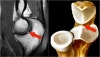
1. Pseudodefect of the capitellum
Pseudodefect of capitellum refers to an abrupt contour change of the posterolateral margin of capitellum on coronal sections and is a potential MRI imaging pitfall giving rise to misinterpretations.
It occurs because the width of the articular surface of the capitellum is non-uniform and gradually tapers posteroinferiorly.
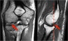
2. Pseudo-loose body
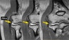
3. Plica
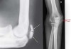
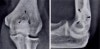
4. Os supratrochlear dorsale
An accessory ossicle of the elbow located in the olecranon fossa of the humerus.
It may become symptomatic due to trauma during elbow extension and as such may require surgical removal.
The differential diagnosis is an intra-articular loose body but the treatment if symptomatic remains the same.
5. Patella cubiti
Patella cubiti is a very rare anomaly of the elbow, presenting as a sesamoid within the distal triceps brachii tendon.
Its exact aetiology is unknown with congenital, developmental and post-traumatic theories postulated.