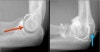
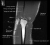
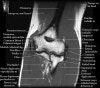
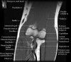
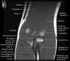
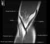
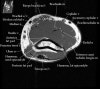
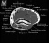
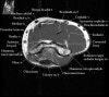
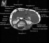
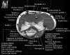
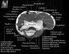
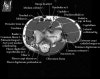
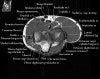
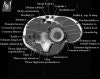
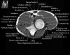
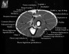
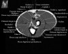
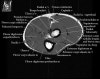
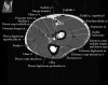
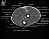
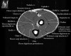
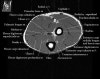
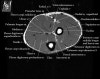
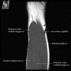
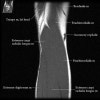
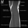
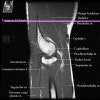
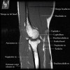
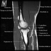
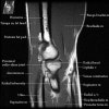
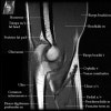
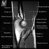
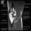
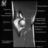
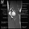
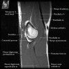
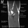
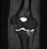
팔꿈치 관절의 MRI 단면 영상
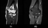
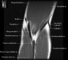
1. Coronal section
1) Collateral ligament
2) Common extensor/flexor tendon group patholgy as well as epicondylitis
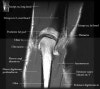
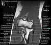
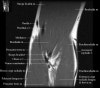
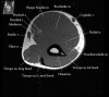
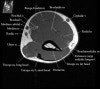
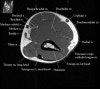
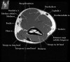
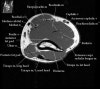
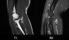
2. Axial section
1) The tendons of the Biceps Brachii and Brachiallis muscles transversely as they insert onto the Radius and Ulna respectively. 2) The distal Triceps tendon is also well evlauated in this plane.
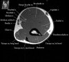
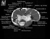
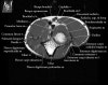

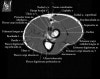
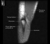
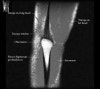
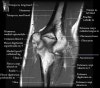
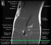
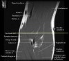
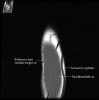
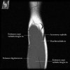
- Sagittal section
1) Biceps Brachii tendon and Brachiallis muscles
2) Radial head for radiographically occult fractures
3) Distal Triceps tendon
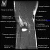
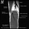
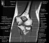
4. MR Arthrography : Useful for evaluation of the collateral ligaments and cartilage surfaces.
5. Elbow Arthrography : UCL pathology in throwers, Osteochondral lesions and repair, Loose bodies