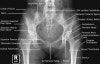
Pelvic X-ray의 Anatomy와 Positioning
- A/P Pelvis
Demonstrates: Acetabulum, pelvic ring, femoral neck, femoral heads
Helpful for: Acetabular fx, Pelvic ring injury, Femoral neck fx, IT fx, hip arthritis, THA, AVN
Position: Supine with feet interally rotated 15°. Center cassette midway between ASIS and pubic symphysis. If feet externally rotated lesser trochanter is visible.
Beam directed perpendicular to plate.

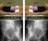
Adult Pelvis AP View(Male)
Adult Pelvis AP View(Female)
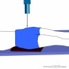
2. Inlet View
Demonstrates: egg-shaped appearance of pelvic ring, sacral promontory should overlap the anterior cortex of the body of the first sacral vertebra
Helpful for: Acetabular fx, Pelvic ring injury, sacral fracture
Position: Supine Center cassette midway between ASIS and pubic symphysis.
Beam directed 40° cephalad
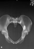
3. Outlet View
Demonstrates: Wedge-shaped appearance of the sacrum when viewed from the front, iliac wings, pubic rami. Top of the symphysis pubis should be at the level of the second sacral body.
Helpful for: Acetabular fx, Pelvic ring injury, sacral fracture
Position: Supine. Center cassette midway between ASIS and pubic symphysis.
Beam directed 45° caudad
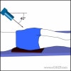
4. Judet Views (obturator oblique and iliac oblique)
Demonstrates: acetabulum
Helpful for: Acetabular fx
Position: Supine on cassette, roll the patient 45 degrees in relation to the x-ray beam. Consider premedication for pain. May be obtained digitally without rolling the patient (Patel NH, J Orthop Trauma 1998;12:59)°
Beam directed perpendicular to cassette.
위로 올라온 측면에서는 Acetabulum의 후면과 Obturator foramen
아래에 놓인 측면에서는 Acetabulum의 전면과 Iliac wing을 잘 관찰