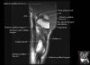
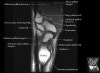
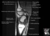
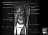
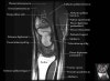
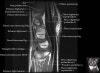
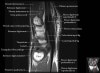
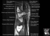
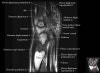
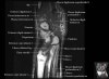
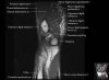
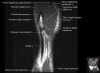
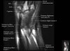
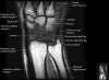
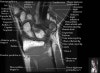
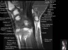
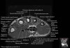
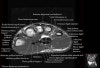
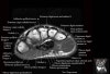
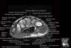
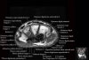
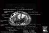
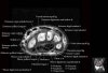
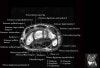
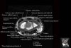
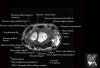
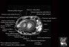
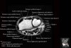
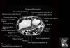
손목 관절의 MRI 단면 영상
- Coronal section
Coronal T1 imaging evaluates bone marrow signal (ex. increased in avascular necrosis) and the relationship of the osseous structures to each other (ex. scapholunate disassociation).
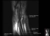
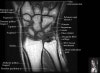
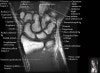

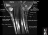
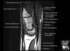
- Axial section
Axial T1 evaluates the tendons of the wrist and carpal tunnel, including the flexor retinaculum.
Axial PD fat suppressed evaluates the tendons of the wrist and carpal tunnel, including the median nerve.
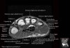
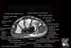
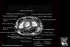
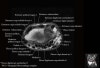
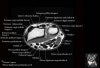
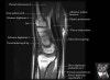
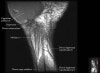
- Sagittal section
Sagittal T1 evaluates the tendons, bone marrow and relationships between the osseous structures.