발목 관절의 MRI anatomy
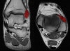
1. Anterior Talofibular ligament
Coronal (left) and axial (right) T1W images assessing the normal normal anterior talofibular ligament.
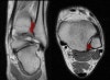
2. Calcaneofibular ligament
Coronal (left) and axial (right) T1W images assessing the normal calcaneofibular ligament.
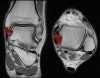
3. Posterior Talofibular ligament
Coronal (left) and axial (right) T1W images assessing the normal posterior talofibular ligament. This ligament is normally less dark than the anterior talofibular ligament.
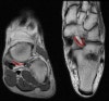
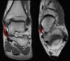
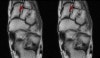
4. Anterior Tibiofibular ligament
Coronal (left) and axial (right) T1W images assessing the distal anterior tibiofibular ligament which is thickened but intact due to a previous injury in this patient.
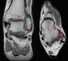
5. Posterior Tibiofibular ligament
Coronal (left) and axial (right) T1W images assessing the normal assessing the distal posterior tibiofibular ligament.
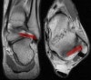
6. Deep deltoid ligament
Coronal (left) and axial (right) T1W images assessing the deep portion of the deltoid ligament.
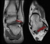
7. Superficial deltoid ligament
Coronal (left) and axial (right) T1W images assessing the superficial portion of the deltoid ligament.
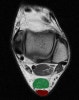
8. Achilles tendon
Axial T1W image assessing the normal Achilles tendon (red). Normally the Achilles tendon is concave or flat on axial imaging.
If it is convex or if it is >6 mm it is abnormal. Incidental accessory soleus muscle (green)
9. Lisfranc ligament
10. Spring ligament
Coronal (left) and axial (right) T1W images assessing the normal spring ligament.