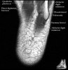
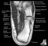
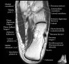
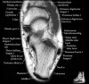
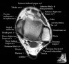
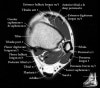
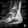
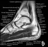
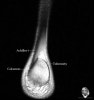
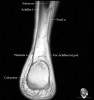
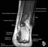
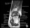
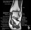
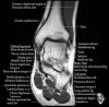
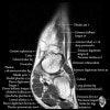
발목 관절의 MRI 단면 영상
- Sagittal section
-
Bones and marrow
-
Joint fluid
-
Talar dome
-
Subtalar joints
-
Achille's tendon
-
Sinus tarsi
-
Plantar fascia
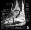
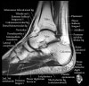
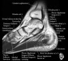
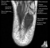
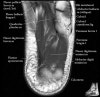
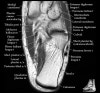
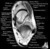
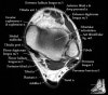
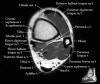
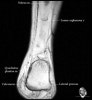
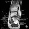
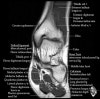
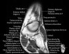
- Coronal section
The coronal oblique T1 sequence follows the tendons of the ankle around the malleolar turns and also evaluates the medial ankle ligaments.
The coronal oblique PD fat suppressed sequence follows the tendons of the ankle around the malleolar turns and is particularly important in evaluation of the Posterior tibialis tendon.
-
Bone and marrow
-
Talar dome
-
Deltoid ligament
-
Tendons in arch
-
Plantar fascia
-
Troubleshoot ligaments
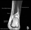
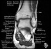
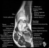
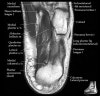
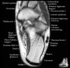
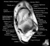
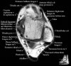
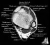
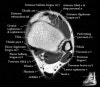
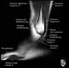
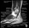
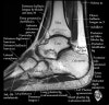
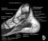
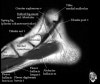
- Axial section
Axial PD fat suppression evaluates the tendons and ligaments of the ankle particularly after acute/subacute injuries. It also is sensitive to talar dome osteochondral defects. Alternatively, a T2 sequence can be used to eliminate magic angle artifact that may occur as the tendons travel around the malleolar turns.
-
Tibiofibular ligaments
-
Lateral ankle ligaments
-
Deltoid and spling ligaments
-
Tendon(Achilles, Medial, Lateral, Anterior)