Knee X-ray에서의 연부조직(Soft tissue) 체크
Horizontal Ray Lateral view는 외상 환자의 연부조직의 관절 삼출액을 관찰하는데 유용하고 특히 지방혈관절증(지방-체액 층)을 알 수 있있습니다.
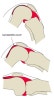
1. Soft tissue signs of Knee effusion(adapted from Weissman and Sledge, 1986, p522 (1))
1) Suprapatellar pouch width greater than 5mm
2) Blurring of posterior aspects of quadriceps tendon
- Increased soft tissue density (fluid density) around anterior femoral condyles (Hoffa's triangle)
4) Widened joint space
5) Bowing of Quadriceps tendon
6) Anterior displacement of patella
7) Bulging of Posterior fat lines
8) Displacement of fabella
- Suprapatellar pouch
1) The knee should be flexed no more that 30 degrees when performing lateral knee radiography.
2) Flexion of the knee greater than 30 degrees can distort/compress the suprapatellar pouch and its adjacent soft tissue structures.
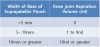
* Estimating the size of Effusions
3. Lipohaemarthrosis
1) Refers to the presence of a blood and fat in a joint.
2) Indicates that there is a fracture that communicates with the knee joint(관절내골절 환자의 35%에서 발생).
- Effusion Cases
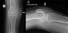
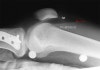
- Very Small
There appears to be a small amount of fluid in the supra-patella pouch (left arrow).
There is also fluid in Hoffa's Triangle (right arrow).
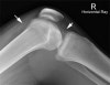
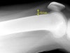
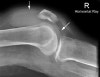
- Small
This patient presented with a history of dislocated right patella.
There is a small amount of fluid in the supra- patella pouch (left arrow) and in Hoffa's Triangle (right arrow).
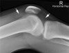
3) Moderate Large
This is evident from the fluid density seen in the supra-patella pouch (left arrow) and in Hoffa's fat pad (right arrow).
A knee effusion indicates an intra-capsular injury (but not necessarily a fracture).
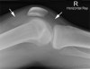
4) Very Large
This elderly patient fell onto her right knee.
She has sustained a fractured patella and has a very large knee joint effusion (arrowed).
* Extra-articular swelling