발의 X-ray anatomy
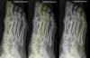
1. Medial column
1) 1st metatarsal
2) Medial cunieform
3) Navicular
4) Talus
2. Middle column
1) 2nd & 3rd metatarsals
2) Middle & lateral cunieforms
3) Navicular
4) Talus
3. Lateral column
1) 4th & 5th metatarsals
2) Cuboid
3) Calcaneus
Lateral column of foot provides majority of mobility and weight bearing in the foot, Significant alterations in foot biomechanics occur with lateral column joint disruption or loss of length.
Note that the cuboid articulates with the bases of the 4th and 5th metatarsals and the calcaneum.
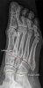
1. Lisfranc joint (forefoot articulation, red line)
2. Chopart joint (hindfoot articulation, yellow line)
To lessen ambiguity, some investigators have suggested that the term "Lisfranc joint complex" should be used to refer to tarsometatarsal articulations and that the term "Lisfranc joint" should be applied to medial articulation involving the first and second metatarsals with the medial and middle cuneiforms.