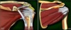
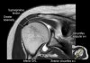
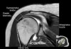
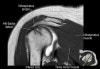
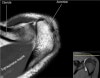
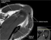
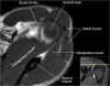
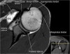
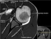
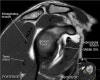
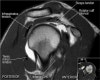
어깨 관절의 MRI 단면영상
- Anatomy
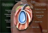
- Coronal section
1) Supraspinatus, Inpraspinatus tendon
2) AC joint
3) Labrum(superior, inferior)
4) Biceps anchor, Inferior GHL, Axillary recess
5) GH joint cartilage
6) Osseous structure
7) Deltoid
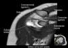
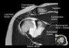
Long head 바깥의 고신호는 Biceps groove의 Normal fluid
Acromion의 하면에 부착하는 저신호 : Deltoid tendon or Corocoacromial ligament일수도 있음
Enthesophyte로 오진가능하나 정상 부착부 소견

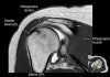
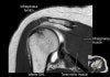
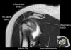
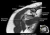
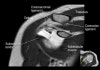
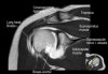
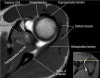
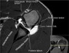
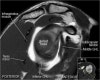
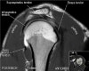
- Axial section
1) Anterior & posterior labrum
2) Subscapularis muscle & tendon
3) Biceps long head tendon
4) GH joint cartilage
5) Osseous structure
6) 관절액 있을때 FSE T2 FS에서 Labrum 선명, 관절액 없을때 FSE PD FS에서 Labrum 선명
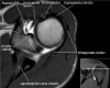
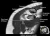
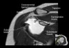
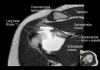
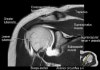
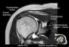
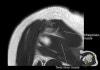
- Sagittal section
1) T1WI : Rotator cuff muscle size, fat infiltration, Biceps tendinosis
2) T2WI : Rotator cuff insertion, edema, Rotator cuff partial or full thickness tear, Bursa fluid collection, Acromial type ,Glenoid fossa, Superior, middle, inferior GHL
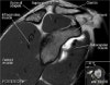
근육 안에 tendon이 들어있다
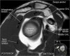
원외부에서 SST와 IST가 겹침